CRIME-LITE APPLICATIONS //
Utilising Crime-lites for The Detection of Challenging Bloodstains
Background
Introduction
Blood is often a critical form of forensic evidence. Its analysis can aid in reconstructing events at a crime scene, identifying individuals, and potentially linking suspects to criminal activity. On light-coloured surfaces, blood is typically easy to detect due to the strong contrast it creates against the background. However, detecting blood on darker substrates presents a greater challenge, as both the blood and the surface absorb significant amounts of visible white light—resulting in minimal contrast when using standard white illumination.
Overcoming the issue
To overcome this limitation, a combination of alternate light sources (ALS) and appropriate filters can be used to reveal blood stains that may be faint or completely invisible to the naked eye. This can assist examiners with locating bloodstains on a surface and can aid in visualising the distribution of the blood on the surface as well.
 |
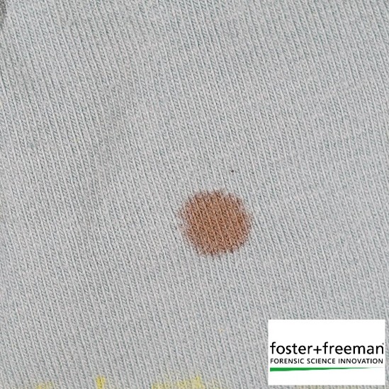 |
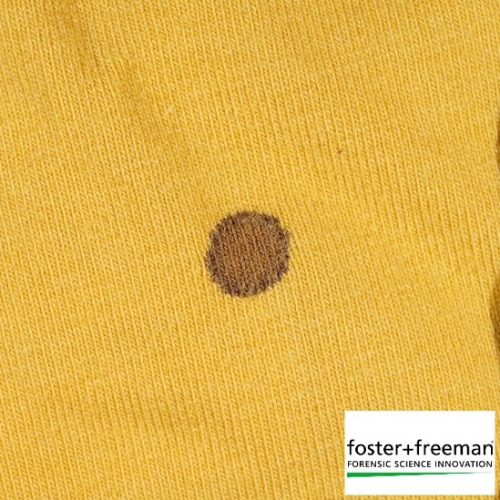
When blood staining is present on a light-coloured surface, it’s often very easily visualised, as shown in the above examples. Because the blood is much darker in colour than the surface, the contrast is visible to the naked eye.
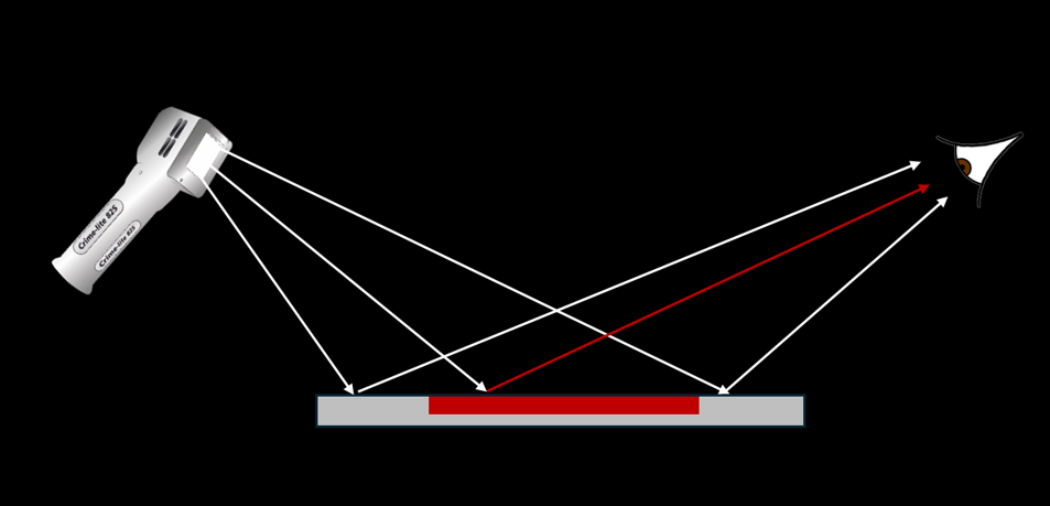
White light is made up of all wavelengths within the visible spectrum. When a surface is illuminated with white light, different colours on the surface absorb and reflect varying amounts of this light. The colours we see as the viewer are those that are reflected to the eye.
This optical principle is useful in for blood detection. Light-coloured substrates typically reflect a high proportion of the incident white light, therefore appearing light to the observer. This then creates contrast with dried blood absorbs which much of the light, resulting in a distinct red-brown appearance. This contrast is what gives visibility to bloodstains and facilitates the location of potential bloodstains during visual searches.
IR Examination
Infrared (IR) light reveals what standard white light can’t. While some surfaces may appear dark to the naked eye because they absorb a high proportion of visible light, they can behave very differently under IR illumination. Depending on the material and the dyes used, these surfaces may actually reflect IR light, appearing much lighter as a result.
This shift in contrast can be really powerful when searching for blood. As blood typically absorbs IR light, stains that are hidden or invisible under white light can become clearly visible against a lightened background in the infrared, making detection quicker, easier, and often far more effective.
 |
 |
| White Illumination |
IR Illumination |
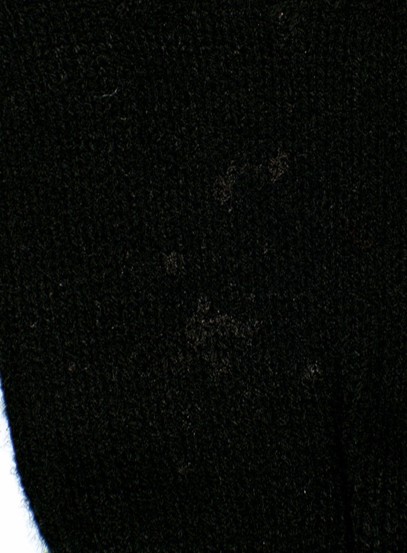
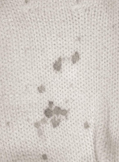
When viewed under standard white light, the dark surface shown here effectively conceals much of the bloodstaining present. However, when illuminated with IR light and observed using a full-spectrum camera and the appropriate filter, the true extent of the blood becomes immediately clear. The use of a full-spectrum camera is crucial, as the human eye is not sensitive to IR wavelengths of light.
Using a modified camera as featured in the Crime-lite® Auto or ML-Pro 2, examiners can harness this capability to locate and document bloodstains on dark or visually complex surfaces. Beyond detection, this method also reveals critical details about the distribution and patterning of the stains—providing valuable context to crime scene examiners.
Fluorescence Examination
Fluorescence is a powerful tool we frequently use when searching for forensic evidence. While blood isn’t typically considered to be fluorescent, this technique can still be highly effective and is often overlooked in blood detection.
In some cases, fluorescence can be induced in the substrate itself, causing it to appear to glow under certain wavelengths of light. This creates strong contrast, as the bloodstains remain dark against the illuminated background. In other scenarios, particularly with aged or degraded samples, it’s possible to stimulate fluorescence in the bloodstains themselves, making them easier to detect.
Whether by enhancing the background or highlighting the stain, fluorescence offers an additional layer of versatility in the search for blood evidence.
Cyanoacrylate Fumed Fingermarks
Traditionally, fumed marks require fluorescent dye stains to stand out. RUVIS simplifies this workflow by revealing high-contrast fingermarks while suppressing complex backgrounds, such as those found on drinks cans, polymer currency, and plastic packaging.
 |
 |
| Blood Fluorescence Examination | Blood 445nm Illumination + 495nm Filter |
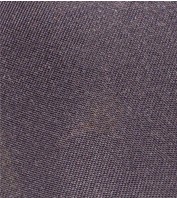
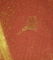
In this example, the bloodstain is completely invisible under white light. However, when examined using fluorescence, the stain itself fluoresces, making it stand out clearly against the background.
This technique can dramatically simplify the detection of otherwise hidden evidence. By enhancing visibility, fluorescence ensures that crucial evidence is not missed during examination, supporting a more thorough investigation.
Contrast Examination
Blood has a strong absorption peak at around 412nm—within the violet region of the spectrum. By illuminating a suspected area with violet light and viewing it through a matched bandpass filter, we isolate how the surface interacts specifically with this wavelength.
Under these conditions, bloodstains often appear significantly darker than the surrounding substrate, creating high contrast and dramatically improving visibility. This targeted lighting technique is a simple yet highly effective way to enhance the detection of blood evidence—especially in cases where stains may otherwise go unnoticed under standard white light.
 |
 |
| Blood Contrast Examination White | Blood Contrast Examination IR |
 |
|
| 410nm Illumination + 415 BP Filter |
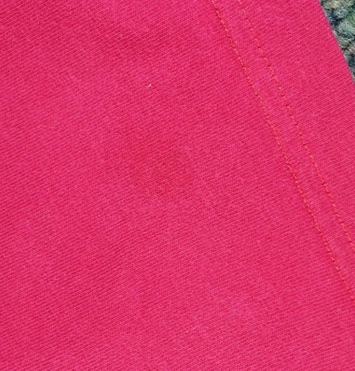
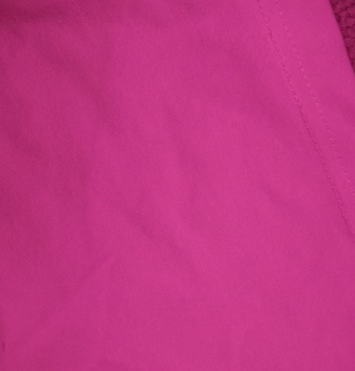
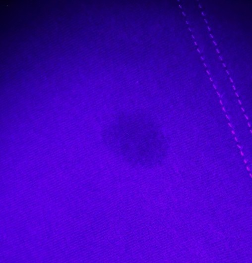
This image shows a small area of dilute blood staining. Under standard white light, the stain is very faint and could easily be overlooked during examination. Even when viewed under infrared (IR) light, no additional contrast is produced, and the stain remains invisible.
However, when the area is illuminated with violet light (around 410nm) and viewed through a matching filter at 415nm, the blood absorbs the light and appears significantly darker than the surrounding surface. This targeted wavelength technique creates high contrast, making the bloodstain clearly visible and easy to detect.
Crime-lite® AUTO
Detect more evidence using the all-in-one forensic search tool
LEARN MORE
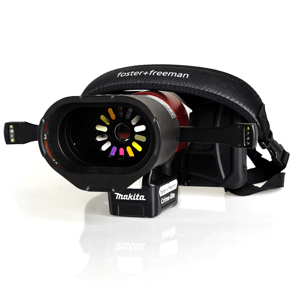
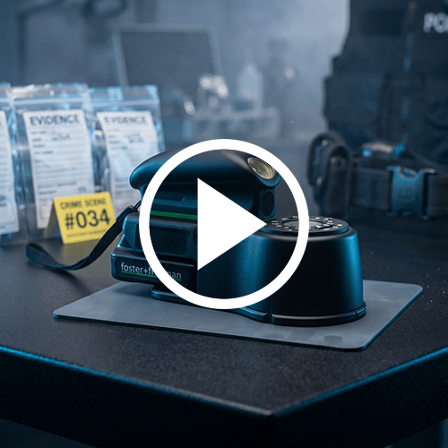
Combining advanced forensic imaging technology with high-intensity multi-spectral illumination, the Crime-lite AUTO is a complete solution to the search, detection, and capture of evidence.
Operated via a simple touchscreen interface, with intelligent filter selection, the Crime-lite AUTO can reveal evidence including fingermarks, body fluids (semen, saliva, and urine, etc.); blood on dark fabrics; fingerprints; gunshot residues; and traces of physical evidence (glass, fibres, hairs, etc.)
- A complete, compact forensic search tool
- All-in-one integrated lights, camera and filters
- Real-time visible, fluorescent, and IR examinations
- Search Beyond the Visible to detect every trace
- Optional laboratory docking system